Morphology
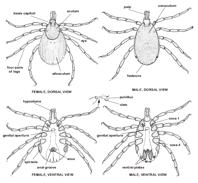
The Ixodidae are characterized by the presence of a tough sclerotised plate on the dorsal body surface, the scutum, covering the entire dorsal body surface in males (sometimes named conscutum), and limited to the anterior approximately one third of the dorsal body region in unfed females, nymphs and larvae. The folded cuticle posterior to the scutum constitutes the alloscutum. Both scutum and alloscutum are covered with numerous small setae. Sexual dimorphism is apparent only in the adult stage. The scutum is the site of attachment of various dorso-ventral body muscles, cheliceral retractor muscles, and many other muscle groups in the Ixodidae. Eyes, if present, are located on the lateral margins of the scutum.
General anatomy of male and female hard ticks. From “Ticks of Domestic Animals in Africa” (Walker et al., 2003) |
Anterior to the scutum, the mouthparts protrude beyond the body and are readily visible dorsally. The mouthparts include the paired chelicerae dorsally and the segmented palps, and ventrally the denticulate hypostome, all mounted on the basis capituli. These structures constitute the capitulum. In all ixodid ticks, the palps consist of 4 segments (=articles), but the tiny sensilla-bearing terminal (4th) segment is recessed in a cavity on the ventral surface of segment III.
Females have a pair of porose areas (= areae porosae) on the dorsal surface of the basis capituli.
Mouthparts of Amblyomma spp., dorsal view |
Ventrally, nymphs and adults bear a pair of spiracular plates (= stigmata) located immediately posterior to the fourth coxae, with the spiracle, a single opening, within each plate.
Pad-like pulvilli occur just proximal to the claws on the tips of the tarsus (last segment of the legs), enabling ticks to climb virtually any surface.
Nymphs and larvae resemble the adults, but lack the external genital pores and porose areas.
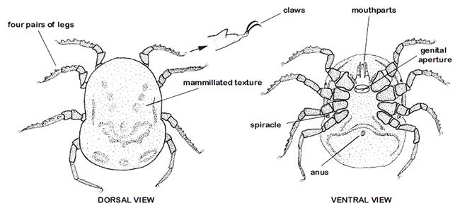
The Argasidae are characterized by a tough leathery integument in all but the larval stage.
The capitulum is recessed ventrally near the anterior end and is not visible dorsally (except in larvae).
Eyes when present are on the lateral surface of the body.
The spiracles or stigmata occur in the supracoxal folds between the coxae of legs III and IV.
Nymphal and adult argasids also bear a pair of tiny pores, coxal pores, representing the openings of the coxal glands, located between the paired coxae of legs I and II. Excess fluid filtered from the bloodmeals they take is excreted via these pores.
External structure of adult argasid ticks (the example is Ornithodoros). From “Ticks of Domestic Animals in Africa” (Walker et al., 2003) |
